- 73.1k views


Use of Ultrasound in ICU
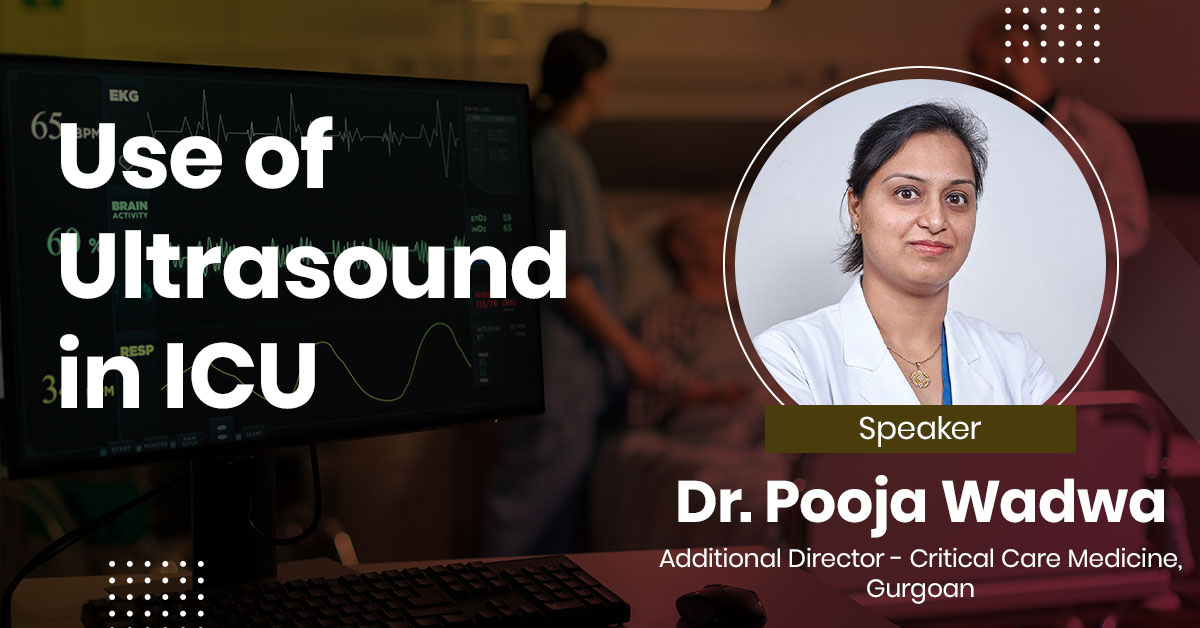
Intensivists who manage critically sick patients frequently employ point-of-care ultrasonography (POCUS), which allows them to correctly and quickly check for a variety of diseases, including pneumothorax, pulmonary edema, hydronephrosis, hemoperitoneum, and deep vein thrombosis, among others. Transesophageal echocardiogram is commonly used in basic and advanced critical care echocardiography to identify the cause of hemodynamic instability in undifferentiated shock and to direct treatment. Studies have shown that respiratory fluctuation of the IVC is unreliable, and the most promising POCUS technique for assessing volume status is monitoring of aortic blood flow velocity following passive leg raising procedures.
About the Speaker

Dr Pooja Wadwa
Additional Director, Critical Care Medicine, ECMO specialist,FMRI,Gurgoan
Upcoming Case Discussions

eIntegrity: Advancing Healthcare for Workers Globally
During the webinar, Mr. Graves will provide insights on - eIntegrity's mission, links with the Royal Colleges in United Kingdom - Extensive range of programmes, and 2022 activity. - He will also highlight the key benefits of eIntegrity courses for healthcare professionals, including world-class e-learning developed by clinicians for clinicians, availability online 24/7, highly engaging and interactive content, suitability for training and professional development, and support for traditional and new learning approaches. About eIntegrity eIntegrity operates independently from Health Education England and is accountable to the eIntegrity Executive Board, which comprises members from the Royal Medical Colleges and Health Education England. By providing high-quality, accessible training and education for healthcare professionals, eIntegrity aims to improve patient care and outcomes worldwide.

Case Based Approach to Upper GI Bleed
Finding the source of the bleeding is essential for the effective management of upper gastrointestinal (GI) bleeding, and if this is done, endoscopic treatment is frequently available. However, because of the bleeding's location or other technical factors, identifying it can be difficult. Consequently, it may be required to employ methods other than endoscopy, including CT angiography. A rare cause of upper gastrointestinal bleeding, duodenal diverticula can be difficult to identify since they sometimes call for specialized endoscopic treatments, like side-viewing endoscope.This case covers the details of first instance of this uncommon syndrome being successfully managed using an upper GI endoscopy using a colonoscope, followed by intravascular coiling.
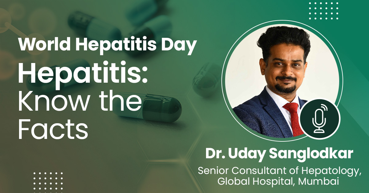
Hepatitis: Know the Facts
Hepatitis is an inflammation of the liver, often caused by viral infections, toxins, or autoimmune diseases. The most common types are Hepatitis A, B, and C, each with different modes of transmission and severity. Hepatitis A is typically spread through contaminated food or water, while Hepatitis B and C are usually transmitted through blood or bodily fluids. Symptoms can include jaundice, fatigue, abdominal pain, and nausea. Chronic Hepatitis B and C can lead to serious complications such as liver cirrhosis or liver cancer. Vaccines are available for Hepatitis A and B, but there is no vaccine for Hepatitis C. Early detection and treatment are crucial for managing and preventing severe liver damage.
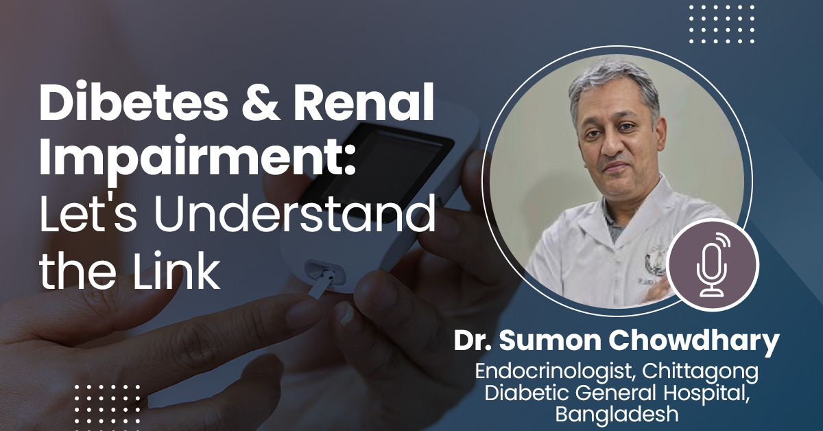
Diabetes and Renal Impairment : Let’s Understand the Link
Individuals with diabetes often develop kidney disease or damage over time. We refer to this kind of kidney disease as diabetic nephropathy. Nephrons in diabetics gradually thicken and get damaged over time. Urine starts to contain protein (albumin) due to nephron leaks. This damage may occur years before kidney disease symptoms appear. When type 2 diabetes develops slowly, kidney damage may already be present in some patients when they are first diagnosed.

Approach to Benign Breast Diseases
The breasts are intricate structures made up of tissue, fat, and glands. Developing a breast lump, cyst, or tumor is a rather typical occurrence. We call this benign breast illness. Even though none of these breast disorders are deadly or malignant, they could raise your chance of getting breast cancer in the future. Even while the majority of breast diseases don't result in cancer, the idea is to get familiar with the feel and appearance of breasts to recognize changes even if the majority of breast diseases are not malignant.
Author Post
Impact
+
Talks
+
webinar
+
no.of registrations
One liner about speaker
Why is speaker relevant?
Dr Pooja Wadwa's Talks on Assimilate
- 7th-November-2022, TIME : 5:00PM - 6:00PM
- 0
Intensivists who manage critically sick patients frequently employ point-of-care ultrasonography (POCUS), which allows them to correctly and quickly check for a variety of diseases, including pneumothorax, pulmonary edema, hydronephrosis, hemoperitoneum, and deep vein thrombosis, among others. Transesophageal echocardiogram is commonly used in basic and advanced critical care echocardiography to identify the cause of hemodynamic instability in undifferentiated shock and to direct treatment. Studies have shown that respiratory fluctuation of the IVC is unreliable, and the most promising POCUS technique for assessing volume status is monitoring of aortic blood flow velocity following passive leg raising procedures.