- 82k views


Cardiovascular Monitoring & Support in Critical Care
Cardiovascular monitoring in critical care involves continuous assessment of vital signs, including heart rate, blood pressure, and cardiac rhythm. Non-invasive techniques such as electrocardiography (ECG) and blood pressure monitoring provide real-time data on cardiac function. Invasive monitoring methods, like arterial catheterization and central venous catheterization, offer more detailed information on hemodynamics and fluid status. Advanced monitoring modalities, such as echocardiography and pulmonary artery catheterization, aid in assessing cardiac function and guiding therapeutic interventions. Supportive measures such as fluid resuscitation, vasopressor therapy, and inotropic support help optimize cardiac output and tissue perfusion. Mechanical ventilation strategies, including positive end-expiratory pressure (PEEP), can improve oxygenation and reduce cardiac workload in critically ill patients.
About the Speaker

Dr. Atchyuth R Gongada
HoD and Sr Consultant Dept of Critical Care and Anaesthesiology Apollo Hospitals, Health city, Visakhapatnam
Dr Atchyuth R Gongada MD FRCA is HoD and Sr Consultant in Dept of Critical Care and Anaesthesiology at Apollo Hospitals, Healtcity, Visakhapatnam.He has done M.B.B.S. from Andhra Medical College and his M.D. (Anesthesia) from Rangaraya Medical College and Obtained fellowship from the Royal College of Anesthesia, U.K. (F.R.C.A.) in 2008. Dr Atchyuth has worked with great people in the field of Anesthesia and intensive care like Dr. Anna Batchelor, Prof. David K.Menon (Founder of Neuro Critical Care Unit, Cambridge). Prof. Arum K.Gupta (Professor of Anaesthesia, Addenbrookes Hospital, Cambridge). Dr. Ian. F.Russell. His specialities are Anesthesiology and Critical Care.
Upcoming Case Discussions
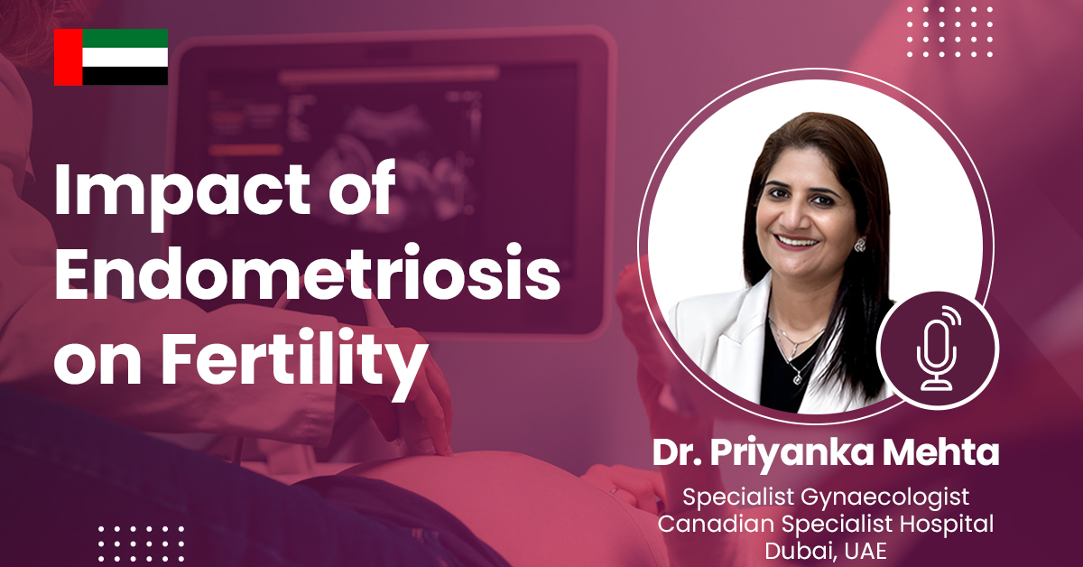
Impact of Endometriosis on Fertility
Endometriosis, a condition where endometrial-like tissue grows outside the uterus, can significantly impact fertility. It causes inflammation, scarring, and adhesions that may distort pelvic anatomy, block fallopian tubes, and impair ovarian function. Endometriosis is also linked to hormonal imbalances and poor egg quality, reducing the chances of conception. Symptoms like chronic pelvic pain and painful intercourse further complicate fertility. Diagnosis often requires laparoscopy, while management includes pain relief, hormonal therapy, and assisted reproductive techniques like IVF. Early intervention with medical or surgical treatment can improve reproductive outcomes, but severe cases may necessitate advanced fertility treatments for conception.
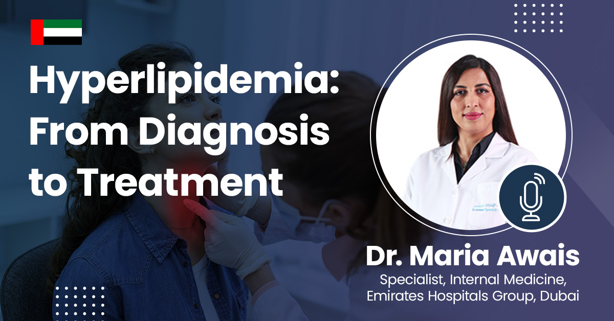
Hyperlipidemia: From Diagnosis to Treatment
Hyperlipidemia is a condition characterized by elevated levels of lipids, such as cholesterol and triglycerides, in the blood, which can increase the risk of cardiovascular diseases. Diagnosis typically involves blood tests measuring lipid profiles, while treatment focuses on lifestyle changes, such as diet and exercise, alongside medications like statins to manage cholesterol levels and reduce cardiovascular risk. Regular monitoring is essential for effective management and prevention of complications.
Acne: Disorders and Treatment Approaches
Acne is a common dermatological condition caused by clogged pores, excess sebum production, bacterial growth, and inflammation. It can manifest as blackheads, whiteheads, papules, pustules, or cysts, often leading to scarring if untreated. Various factors, including hormonal changes, diet, stress, and genetics, influence its severity. Treatment approaches range from topical and oral medications, such as retinoids, antibiotics, and hormonal therapy, to advanced procedures like chemical peels and laser therapy. A personalized skincare regimen, along with lifestyle modifications, plays a crucial role in managing and preventing acne.
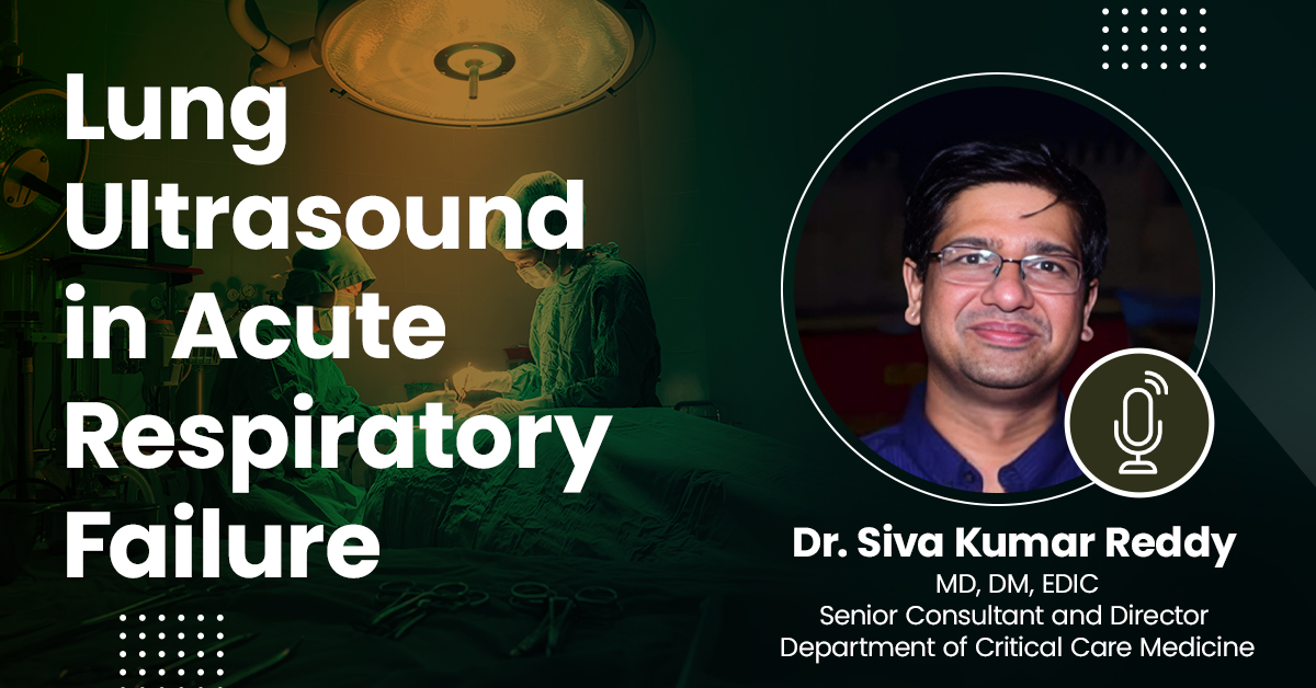
Lung Ultrasound in Acute Respiratory Failure
Lung ultrasound has emerged as a vital, non-invasive tool in the rapid assessment of acute respiratory failure. It provides real-time imaging to differentiate conditions like pneumonia, pulmonary edema, pneumothorax, and pleural effusion with high accuracy. Compared to traditional chest X-rays, lung ultrasound offers superior sensitivity, especially in critically ill patients where bedside evaluation is crucial. Its ability to guide immediate clinical decisions improves patient outcomes and reduces unnecessary radiation exposure. With standardized protocols like the BLUE (Bedside Lung Ultrasound in Emergency) protocol, it enhances diagnostic efficiency in emergency and ICU settings.
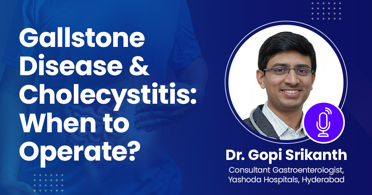
Gallstone Disease & Cholecystitis: When to Operate?
iGallstone disease and cholecystitis are common conditions requiring careful evaluation to determine the need for surgery. Symptomatic gallstones causing recurrent pain, nausea, or complications like cholecystitis often necessitate cholecystectomy. Acute cholecystitis, characterized by inflammation, fever, and right upper quadrant pain, typically requires early surgical intervention to prevent complications like perforation or sepsis. In high-risk patients, conservative management with antibiotics and drainage may be considered. Elective surgery is recommended for asymptomatic patients with high-risk factors, such as large gallstones or gallbladder polyps, to prevent future complications.
Author Post
Impact
+
Talks
+
webinar
+
no.of registrations
One liner about speaker
Why is speaker relevant?
Dr. Atchyuth R Gongada's Talks on Assimilate
- 20th-February-2024, TIME : 05:00PM - 06:00PM
- 0
Cardiovascular monitoring in critical care involves continuous assessment of vital signs, including heart rate, blood pressure, and cardiac rhythm. Non-invasive techniques such as electrocardiography (ECG) and blood pressure monitoring provide real-time data on cardiac function. Invasive monitoring methods, like arterial catheterization and central venous catheterization, offer more detailed information on hemodynamics and fluid status. Advanced monitoring modalities, such as echocardiography and pulmonary artery catheterization, aid in assessing cardiac function and guiding therapeutic interventions. Supportive measures such as fluid resuscitation, vasopressor therapy, and inotropic support help optimize cardiac output and tissue perfusion. Mechanical ventilation strategies, including positive end-expiratory pressure (PEEP), can improve oxygenation and reduce cardiac workload in critically ill patients.